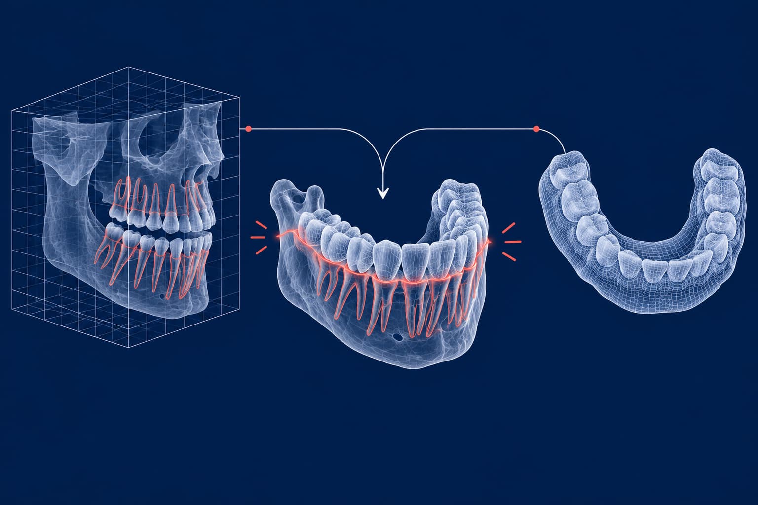
A CBCT scan adds what an intraoral scan cannot see: roots, bone and impacted teeth. An intraoral scan adds what CBCT renders poorly: crisp crown surfaces and gingival margins. The digital workflow merges the two. You export CBCT as DICOM, segment it into an STL surface, register it to your intraoral scan, and design the appliance or guide on the combined model.
The merge is only as good as its weakest step, and the weakest step is usually segmentation, not the scanner. The evidence also says you should not take a CBCT by default. Here is how the pipeline works, where it loses accuracy, and what to send your lab.
| Stage | Input | Output | Main accuracy risk |
|---|---|---|---|
| Justify and acquire | Clinical question | CBCT volume (DICOM) | Field of view larger than needed |
| Segment | DICOM | Bone and tooth STL | Threshold choice, metal artifacts |
| Register | CBCT STL plus intraoral scan | One fused model | Few or poorly chosen landmarks |
| Design | Fused model | Guide, framework or appliance STL | Safety margins ignored |
| Hand off | STL/PLY, sometimes DICOM | Lab-ready case | Unclear file ownership, patient data |
When CBCT belongs in an orthodontic workflow
The European SEDENTEXCT guidelines state that no scientifically valid evidence supports large-volume CBCT at any stage of routine orthodontic treatment, so the panel could not recommend it as a standard method of diagnosis and treatment planning. It may be justified for complex skeletal abnormalities, particularly combined orthodontic and surgical cases.
For an impacted tooth, SEDENTEXCT allows CBCT when lower-dose conventional radiography cannot answer the question, including possible resorption of an adjacent tooth. It asks you to choose the smallest volume that fits the clinical situation.
Dose is the reason. SEDENTEXCT reports effective doses in a 10-year-old phantom of 16–214 μSv for dento-alveolar CBCT and 114–282 μSv for craniofacial CBCT. For an adolescent phantom the ranges are 18–70 μSv and 81–216 μSv. The guideline notes that dose depends on the unit and exposure settings, above all the field of view. The same document stresses that justification matters most in children.
A practical rule follows from this. If the question is local (an impacted canine, a suspected resorption, a bone-anchored device), a small field of view is the better request. If the question is only tooth movement or model analysis, a surface scan already contains what you need; our guide to digital model analysis in orthodontics explains why surface scans are the safer source than segmented CBCT.
SEDENTEXCT also states that CBCT is not normally indicated for planning temporary anchorage devices (TADs). That sets a limit on the guided miniscrew workflow described below: you do not order a CBCT only to place a miniscrew, but if you already have one for a justified reason, you can reuse it for guide design.
From DICOM to STL: where the first errors enter
A CBCT study is a stack of grey-value slices stored as DICOM. The DICOM standard wraps each image with a header that carries identifying information, which matters for the handoff section below. Your design software or a segmentation tool applies a grey-value threshold to separate bone and teeth from soft tissue, builds a 3D surface, and exports it as STL.
That threshold is a decision, not a default. In a 2025 study in the Journal of Prosthetic Dentistry, researchers generated STL bone models from CBCT scans at seven thresholds (250–1750 HU) and aligned intraoral scans to each. Threshold significantly changed the result (P<.001 on all axes). Deviations along the apicocoronal (height) axis reached −0.13 to −0.66 mm, much larger than along the left–right axis (0.01 to 0.03 mm).
A height error of that size is not trivial when a guide has to steer a miniscrew between two roots. If your lab or software vendor segments for you, ask which threshold they use and whether it is adjusted per case.
Another study of 36 patients compared default and manual segmentation. The mean deviation between surface scan and CBCT model was 0.54 mm overall, with 0.69 mm for default segmentation and 0.4 mm for manual segmentation. It also found that the user and the number of restorations affected the result.
Metal is the enemy of clean segmentation
Metal in the field of view causes streaking artifacts that obscure tooth surfaces and complicate segmentation. Brackets, bands and crowns all qualify. Some registration studies exclude patients with fixed appliances for that reason. The consequence for you is about sequence: where the clinical timeline allows, acquire the CBCT before bonding, and take the intraoral scan on clean teeth.
Merging CBCT with the intraoral scan
Fusion works because the two datasets complement each other. A published clear-aligner workflow describes the typical steps. First, align the models using at least three matching anatomical landmarks. Then refine with best-fit alignment. Finally, merge the models, taking crown detail from the intraoral scan and roots from the CBCT.
Landmarks versus surfaces
Many programmes also offer surface-based registration using an iterative closest point (ICP) algorithm, which repeatedly minimises the distance between selected tooth surfaces. A recent registration study compared the two approaches:
- Three-point registration produced deviations of 257.0 μm; three-surface registration produced 479.3 μm.
- Raising the number of registration points from two to four cut deviations from 375.4 to 228.2 μm. Going beyond four gave no significant further improvement.
- Higher CBCT image quality lowered deviations from 314.4 to 237.6 μm.
The authors concluded that clinicians should prioritise point-based registration. The decision rule is simple: pick four well-spread landmarks on stable tooth surfaces (cusp tips, not restored areas), and spend the effort on image quality instead of adding more points.
How accurate is accurate enough?
An automatic registration method reported a mean error of 0.234 mm, below the 0.3 mm voxel size of the CBCT it used. A study using a 0.39 mm voxel found that digital dentition models deviated from the reference by less than one voxel and judged this clinically acceptable. The takeaway is that registration error is normally comparable to voxel size. Anything larger usually points to a segmentation or artifact problem, not to the algorithm.
Automation is closing the time gap. In a validation of an AI-based registration tool on 31 scans, automatic registration took 51.4 ± 17.2 seconds, against 274.7 ± 100.7 seconds for semi-automated and 840 ± 168.9 seconds for manual registration. A separate 2026 study of 45 patients found registration error rising with artifact burden for both AI-assisted and point-based methods, with AI-assisted registration consistently lower. Always inspect the overlay at the roots and bone margin before you approve a design, whichever method you use.
What the fused model makes possible
Once the model is fused, the lab can design things that a surface scan alone cannot support.
Miniscrew insertion guides. The published workflow runs as follows: plan the miniscrew site and direction on the fused data, design the guide, export it as STL and print it, typically by stereolithography. A systematic review of 11 studies and 652 miniscrews concluded that static computer-assisted guidance improved the safety and accuracy of insertion over freehand and radiographic-guide methods. Root damage was significantly lower (OR 0.11).
A clinical study placed 35 mini-implants in 15 orthodontic patients who needed a CBCT for other reasons, such as impacted teeth. Guides designed with CBCT information showed no significant angular difference between planned and achieved position, while guides designed without CBCT were less accurate in inclination.
For anterior-palate guides, a 2025 study found the full-arch design more accurate than the skeletonised design, and recommended building a safety distance into the virtual plan to absorb insertion deviation. Our guide on 3D-printed surgical guides for orthodontic TAD placement covers the lab side.
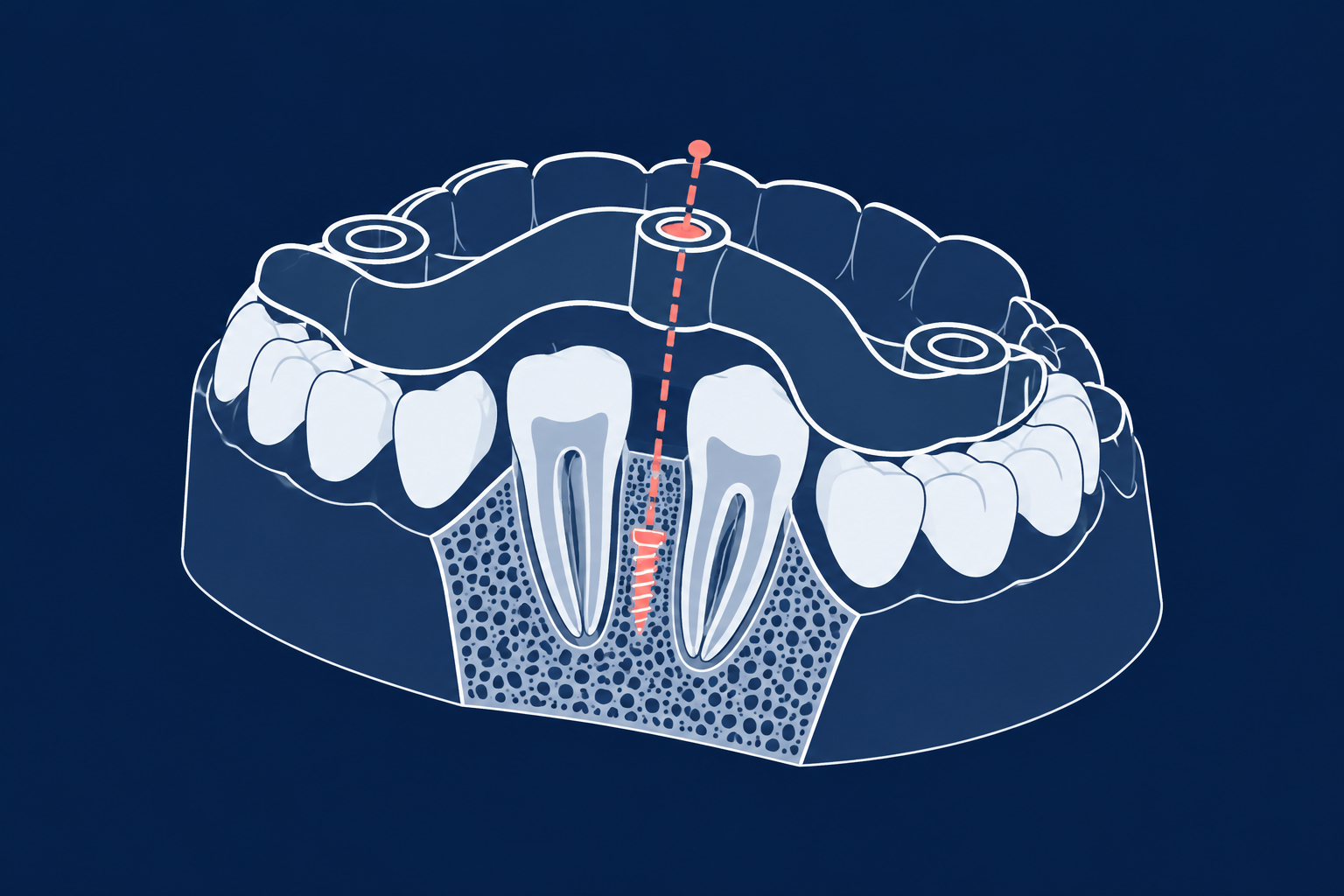
Impacted canines. A case report of two palatally impacted canines used CBCT and intraoral scans to locate the tooth and design a device for exposure and traction. This is the setting where the CBCT is justified in its own right and the digital model adds a design path.
Bone-anchored expanders. Printed MARPE frameworks are designed on CBCT plus scan. See our overview of 3D-printed palatal expanders for types and evidence.
Where it does not help yet. A 2024 scoping review found that CBCT is broadly supported for dental anomalies, temporomandibular joint disorders and craniofacial malformations, but that CBCT upper-airway analysis needs further validation. Do not treat an airway volume number as a treatment target.
Handing CBCT data to the lab
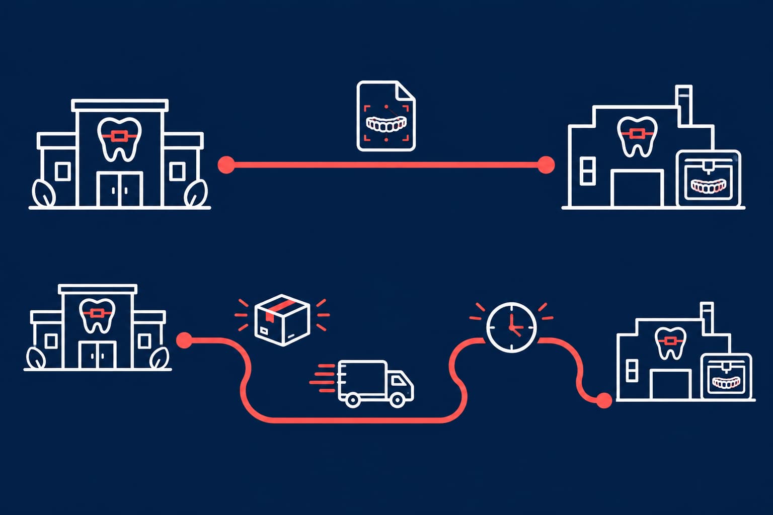
Surface scans travel as STL or PLY. Our article on STL files in orthodontics covers format choices, and open versus closed digital systems explains why open exports matter. CBCT is different, because the raw data is DICOM. Whether a lab accepts DICOM varies, so confirm before the patient is scanned.
Three questions to settle with your lab:
- Who segments? Given the threshold effect above, agree whether you send the raw DICOM or a segmented STL, and who verifies the registration.
- What is the file size? A DICOM study can span hundreds of slice files. One published CBCT study converted a 250 MB DICOM folder into an STL of about 100 MB. That is a single example, not a rule, since size depends on the field of view and voxel size.
- How is patient data protected? DICOM headers carry patient identity. The DICOM standard defines confidentiality profiles that remove or replace identifying attributes. Under the GDPR, health data is a special category, and the EU Court of Justice has noted that processing requires a lawful condition and appropriate security measures, including pseudonymisation or encryption where appropriate. Pseudonymised data is still personal data, so a data-processing agreement with the lab still applies. Ask your data-protection officer to confirm the rules that apply in your country.
Sending only the segmented surface your lab needs is a sensible way to reduce exposure, as long as segmentation quality has been agreed first.
Common mistakes to avoid
- Ordering a large field of view for a local question. Request the smallest volume that answers it.
- Accepting default segmentation. Check the threshold and the result at the root surfaces.
- Registering on restored or banded teeth. Choose natural, artifact-free surfaces for landmarks.
- Skipping the visual check. An automated overlay still needs your eyes at the roots.
- Planning to the millimetre. Guide studies recommend a safety margin.
- Sending identifiable DICOM by ordinary email. Use a secure channel and agree the data terms first.
Your next CBCT case will go faster if the decisions are made before the scan: the clinical question, the field of view, the segmentation owner and the file format. Before you book the scan, read how a digital case moves from scan to appliance, and ask NordicDens which files your case needs at the design stage.